T-cell engagers against a peptide-MHC tumour target
T-cell engagers can only attack what they can see, and what they can see is the cell surface. Fewer than 15% of cellular proteins go there. The other 85% are intracellular, and by the usual logic unreachable. That 85% includes most of the proteins that actually distinguish a cancer cell from a healthy one.
They are not quite unreachable. Every cell continuously chops up its internal proteins and displays the fragments on MHC class I, which is how the immune system audits what is going on inside. So an intracellular tumour antigen does reach the surface, as a short peptide sitting in a groove. Building a T-cell engager that targets one of those would open the other 85% of the proteome.
The difficulty is specificity, and it is severe. An MHC molecule presenting the wrong peptide looks almost exactly like one presenting the right peptide: the same large protein, the same fold, differing in a handful of side chains pointing out of a groove. A binder that latches onto the MHC rather than its cargo will find that cargo on every cell in the body.
Why antibodies rather than engineered T-cell receptors
This is the strategic choice underneath the work, and it is a choice about where the engineering effort goes.
T-cell receptors are the natural readers of peptide-MHC, but their natural affinities are far too weak for a therapeutic, so soluble TCR-based molecules have to be extensively affinity matured. That maturation is where the trouble starts: pushing affinity up tends to push binding towards the invariant MHC framework, and molecules emerge that bind well and no longer care much which peptide is being presented.
Antibodies come at it from the other end. They bind pMHC in the nanomolar-to-picomolar range to begin with, so far less engineering is needed, so there is far less opportunity to drift into peptide-independent binding. The bet is that starting with enough affinity is a better route to specificity than engineering affinity in afterwards.
What was built
The target is MAGE-A4, a cancer-testis antigen, expressed in tumours and, among normal adult tissues, essentially only in testis, which is immune-privileged. Intracellular, so reachable only as pMHC.
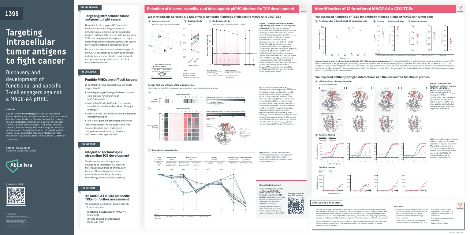
Discovery produced 45 MAGE-A4-pMHC antibodies, from which 6 were taken forward across 4 clonal families, spanning affinities from 7 nM to 15 µM. Even at the panel stage they were counter-screened against 19 unrelated pMHCs.
Those 6 were then paired against 19 CD3-binding antibodies drawn from the functionally clustered CD3 panel, producing over 200 bispecifics in a 1×1 format. Twelve were selected for detailed assessment, spanning roughly four orders of magnitude in potency, and one of those, the lead, carried forward.
What the lead does
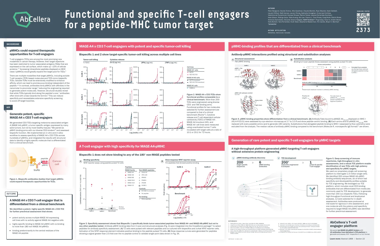
The specificity screen is the result that matters, because it is the one the whole approach turns on. Across a panel of more than 180 non-MAGE peptides, the lead bispecific bound none of them. Its binding is restricted to MAGE-A4 and MAGE-A8 pMHC.
A strong result, and worth reading precisely: restricted to two related antigens, not to one. MAGE-A8 cross-reactivity is real and is the kind of thing that has to be carried forward into preclinical work rather than rounded away.
Functionally, the lead kills around 75% of MAGE-A4-positive cells at the top of the dose range, in two independent lines, with dose-dependent IL-2, IFNγ and TNFα release. Against MAGE-A4-negative lines, and against a MAGE-A4 knockout, there is no activity and minimal cytokine response. The knockout is the control that carries the argument: it holds the cell line constant and removes only the antigen, so the killing is demonstrably antigen-driven rather than a property of those particular cells.
Why it is specific
Cryo-EM structures of the antibody bound to its target answer the mechanistic question directly, and the answer is the one you would want.
The binding is peptide-centric: the antibody contacts predominantly the central residues of the peptide rather than the surrounding MHC framework. Different antibodies in the panel approach at quite different orientations while still reading the peptide, which suggests peptide-centric binding is a repeatable outcome of this discovery route rather than one lucky molecule.
The critical contact is arginine at position 6, whose side chain projects fully out into solvent, making it the single most readable feature the peptide offers. A positional scan across all ten peptide positions confirms Arg 6 as the dominant determinant, and separates the lead’s recognition profile from a clinical benchmark molecule across the full set of positions.
So the structural evidence and the functional evidence agree: this molecule discriminates on the peptide, which is precisely the failure mode that has limited affinity-matured TCR approaches.
Contribution
A supporting author on both posters (25th of 31 at SITC 2023, 23rd of 35 at AACR 2024). It is a large, mostly experimental programme, spanning antibody discovery, cell-killing assays, cytokine panels and cryo-EM. My own line of work connects through the CD3 binders paired in here, which come from the functionally clustered panel.
Where this sits
Two presentations of one programme at successive stages: SITC 2023 covers discovery and the selection of twelve candidates; AACR 2024 covers the deep characterisation of the lead, and cites the CD3-selection work as its front end. I have given them one page rather than two, because they are one body of work and splitting them would duplicate the science while halving the story.
The posters
 Click to enlarge
  Click to enlarge